Poster Presentation Australasian Melanoma Conference 2026
Conjunctival ultraviolet autofluorescence as a potential ocular biomarker of cumulative UV exposure in skin cancer risk assessment: a pilot study (#410)
Background
Skin cancer, including melanoma and keratinocyte carcinomas, is the most commonly diagnosed malignancy in Australia, with ultraviolet (UV) radiation driving up to 95% of cases. Current risk stratification relies on self-reported behavioural and phenotypic factors, which are prone to recall bias and fail to capture biological UV burden at the tissue level. Conjunctival ultraviolet autofluorescence (CUVAF) is a rapid, non-invasive ocular imaging technique that quantifies UV-induced conjunctival damage within seconds (Figure 1). This pilot study assessed the feasibility of clinic-based CUVAF imaging and explored its distribution and correlates in individuals with and without skin cancer.
Methods
In this cross-sectional pilot study, 24 participants were recruited: 15 with confirmed skin cancer (keratinocyte carcinoma n=12; melanoma n=3) and 9 controls. Bilateral nasal and temporal conjunctival images were captured using a handheld device designed and developed by our team. Participants completed a standardised questionnaire capturing demographics, sun exposure, sun protection behaviours and skin cancer history. Given the sample size, associations were explored descriptively using Spearman correlations without adjustment for confounders.
Results
The skin cancer cohort (mean age 67±10 years; 8 male) included keratinocyte carcinomas (n=12) and melanoma (n=3). Controls (mean age 46±14 years; 2 male) had no skin cancer history. CUVAF was detected in 11/14 evaluable skin cancer patients (3/3 melanoma; 8/11 keratinocyte carcinoma) and 4/9 controls. Self-reported outdoor exposure showed no association with CUVAF (r=0.19, p=0.53), highlighting the limitations of questionnaire-based UV assessment.
Conclusions
Handheld CUVAF imaging was feasible in a skin cancer clinic and CUVAF was more frequently detected in skin cancer patients than controls. These findings support further investigation of CUVAF as a rapid, objective measure of cumulative UV burden that could complement existing risk assessment tools. Larger prospective studies are needed to determine whether CUVAF can improve risk stratification and identify high-risk patients.
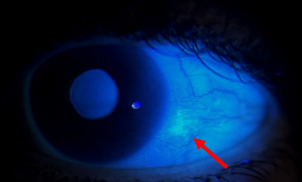
Figure 1. Conjunctival ultraviolet autofluorescence (CUVAF) imaging demonstrating a focal area of UV-associated conjunctival fluorescence, indicative of cumulative UV damage (arrow).