Poster Presentation Australasian Melanoma Conference 2026
Melanoma of Unknown Primary with Cutaneous Regression and Poliosis: A Diagnostic Challenge (#437)
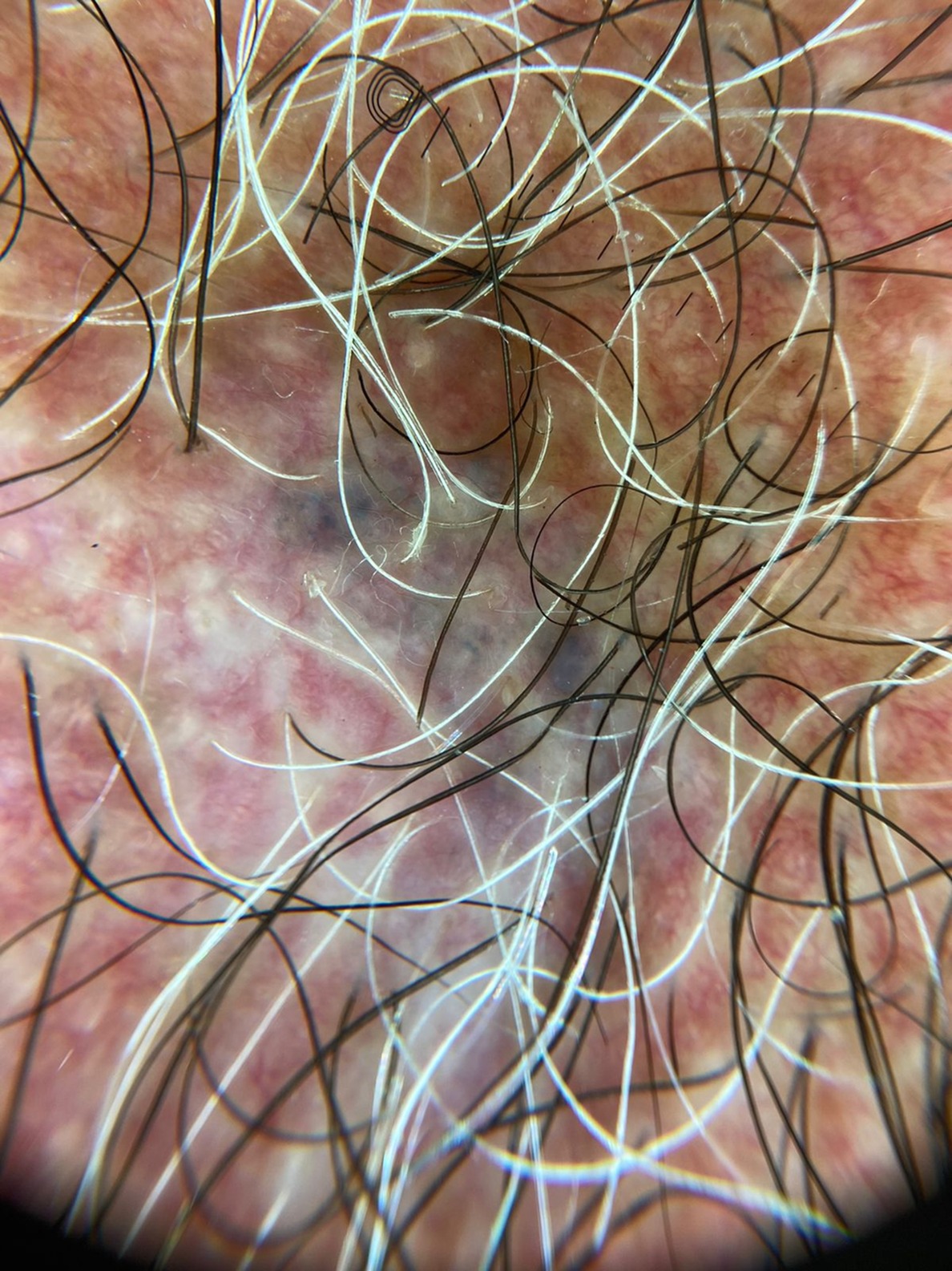
Background/Aim: Melanoma of unknown primary (MUP) represents a significant diagnostic challenge, particularly when regression obscures the primary lesion. Circumscribed poliosis, resulting from T-cell–mediated melanocyte destruction, is a rare but potentially relevant clinical finding reported in association with melanoma. We report a case of metastatic melanoma presenting with neurological deficits and poliosis in the setting of a regressed cutaneous lesion.
Methods: A 39-year-old man presented with progressive right-sided hemiparesis over three months. Brain magnetic resonance imaging revealed a left frontoparietal space-occupying lesion with associated vasogenic edema, suggestive of neoplasia. Systemic workup failed to identify a primary tumor, prompting dermatologic evaluation.
Results: The patient reported a longstanding lesion on the sternum, present for several years, with slight interval growth and a single episode of bleeding following manipulation, without associated pain or pruritus. Dermatologic examination revealed an approximately 3 cm macule in the same location, with subtly hyperpigmented borders and a pale, shiny center. Dermoscopy demonstrated an erythematous background, irregular telangiectasias, peripheral homogeneous blue areas, and central regression structures. The patient also reported recent onset of poliosis involving the beard, chest hair, and scalp. Neuroimaging demonstrated a multilobulated lesion in the left perirolandic region with contrast enhancement, vasogenic edema, and hemorrhagic components, including areas of blood product degradation, associated with multiple satellite lesions and an additional contralateral nodular lesion, highly suggestive of metastatic disease. Incisional and subsequent excisional biopsies of the cutaneous lesion showed no evidence of malignancy, revealing dermal scarring, fibroelastotic proliferation, and xanthogranulomatous inflammation, consistent with regression. Two months later, surgical resection of the brain lesion confirmed metastatic melanoma, with immunohistochemical positivity for S100, Melan-A, and HMB45.
Conclusions: This case highlights the diagnostic complexity of melanoma with occult or regression-masked primary lesions. Circumscribed poliosis, although uncommon, may represent an important clinical clue. Regressed cutaneous lesions may mimic benign entities both clinically and histopathologically, representing a significant diagnostic pitfall. Recognition of these findings is essential to ensure timely diagnosis and appropriate oncologic management.