Poster Presentation Australasian Melanoma Conference 2026
The genomic and transcriptomic landscape of skin adjacent to a prior melanoma is distinct from clinically similar skin without an adjacent melanoma (#419)
Background
Normal skin has a high somatic mutation burden, yet this does not explain where a melanoma will form. Potentially, mutated “tumour-competent” melanocytes are kept in check by a sufficiently functional microenvironment, while a dysfunctional cellular community allows proliferation. The PhotoMelanoma Study aimed to determine the genomic architecture of the microenvironment that favours melanoma formation.
Method
We assessed biopsies of photodamaged skin adjacent to a melanoma excision, photodamaged skin 5cm away from the excision (“proximal”), and distant photoprotected skin from the same individual in 19 participants. Biopsies were assessed for hotspot mutations via droplet digital PCR; somatic mutation burden and signature via deep panel sequencing of 300+ cancer-related genes (PanelSeq), and global methylation profiling of 900K loci. Single cell and spatial morphological transcriptomics were performed on a subset of samples.
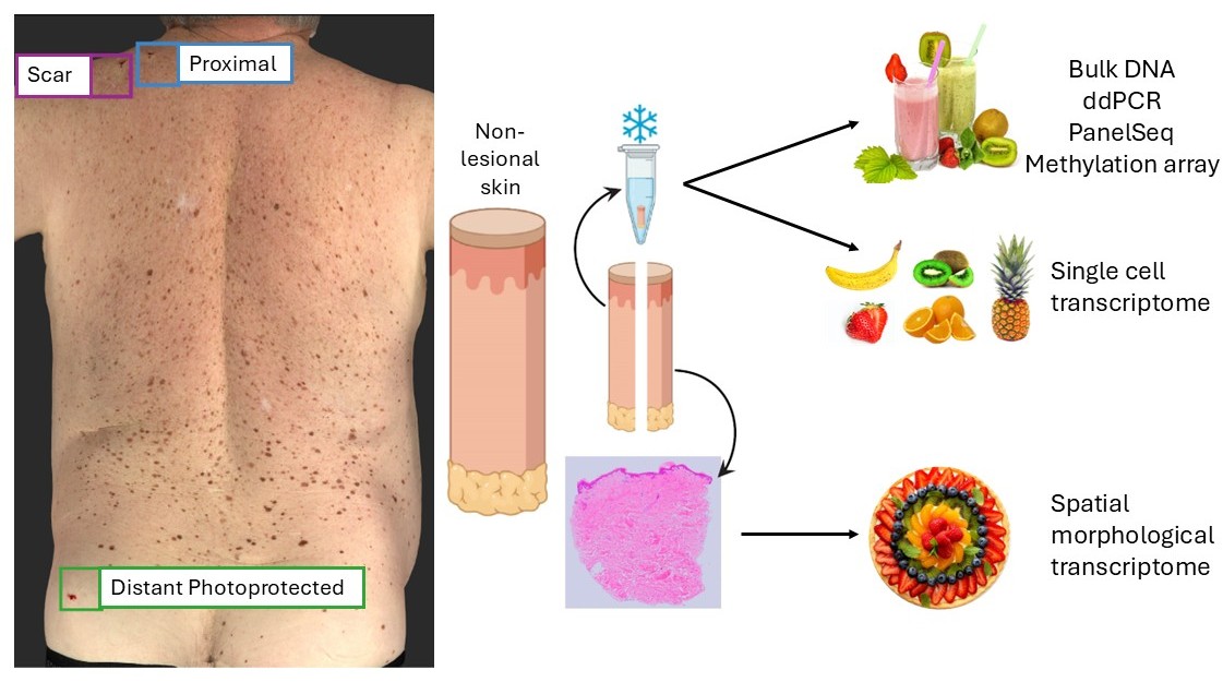
Results
BRAF V600 and TERT promoter driver mutations were detected, with BRAF V600E enriched at scar sites compared to proximal and distant sites (78% vs 52%, respectively; p=0.037). PanelSeq showed UV-related mutation signatures (SBS7) at similar levels across photodamaged sites, whereas APOBEC-related signature SBS2 was enriched (67% vs 29%, respectively; p=0.025) at the scar-adjacent site only.
Interestingly, even within a small region of the skin, there are notable differences in global DNA methylation and cell type-specific gene expression, with differential methylation and expression in tumour suppressors and MAPK signaling genes. There were also significant differences in the HOX gene family, known to influence naevus dermoscopic types and melanoma formation at different body sites, across keratinocytes and fibroblasts.
Conclusions
In sum, there are multiple differences in the genomic and transcriptomic landscape of melanoma-adjacent skin and visually-similar proximal skin, which may point to an increasing loss of the cellular microenvironment’s ability to regulate itself. These data are part of a comprehensive genomic profile to identify underlying causes of melanoma development, to aid development of personalized detection and treatment strategies for melanoma.